The Internal Structure of the Eye
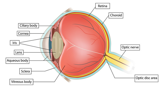
After the Iris and inside the Sclera is the Inner Structure of the eye. This includes
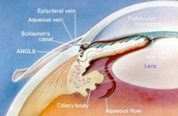
Aqueous Humour
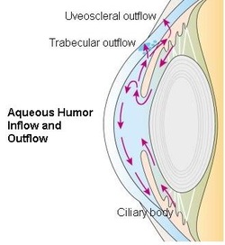
A clear, watery fluid (produced by the Ciliary cody) that fills the area between the Lens and the Cornea. It supplies nutrients that nourishes the Cornea and Lens and maintains the convex shape of the Cornea along with refracting light onto the pupil.
Vitreous Humour
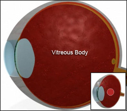
A transparent, jelly-like substance that forms the main bulk of the eyeball. While it maintains the shape of the eye, it also refracts light onto the Retina.
Lens
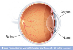
The Lens is a transparent, round, biconvex structure. It is elastic so that its curvative can be changed to adjust its refractive or fucusing power. It's responsible for focusing light onto the Retina.
Suspensory Ligament
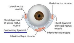
A tissue that joins the lens to the ciliary body. This hold the Lens in place.
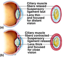
Ciliary Body
A circular band of tissues at the front of the Choroid that supports the Lens. It contains Ciliary Muscles that adjusts the curative of the Lens.
Ciliary Muscles determines the shape of the Lens. When the muscle contracts, the lens is rounder, thicker, the focal length decreases and more convex and light rays from a near object are sharply focused on the Retina. When the muscle relaxes, the Lens is flatter, the focal length increases and less convex and the light rays from a distant object are sharply focused onto the Retina. This can be described as Antagonistic (when one sewt contracts, the other relaxes)
Ciliary Muscles determines the shape of the Lens. When the muscle contracts, the lens is rounder, thicker, the focal length decreases and more convex and light rays from a near object are sharply focused on the Retina. When the muscle relaxes, the Lens is flatter, the focal length increases and less convex and the light rays from a distant object are sharply focused onto the Retina. This can be described as Antagonistic (when one sewt contracts, the other relaxes)
Choroid
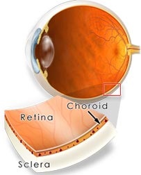
Black-pigmented layer under the Sclera that prevents the internal reflection of light rays. Filled with blood capillaries, It is rich in blood vessels that bring oxygen and nutrients to nourish the eyeball. The Choriod is modified to form the Iris and the Ciliary Body at the front of the eye.
Retina
The inner-most, light sensitive layer of the eyeball, on which images are formed.
They contain Photoreceptors which are light-sensitive cells.
Photoreceptors
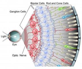
- Rods are responsible to detect light in dim light (black and white) enabling night vision. They contain a pigment in rods called Rhodospin which is light sensitive. If exposed to bright light then it will be bleached. Rhodospin enables a person to see in the dark.
- Cones are responsible to detect coloured light. There are green, blue and red cones. Red is used to respond to long wavelengths. Blue is used to respond to short wavelengths and Green is used to respond to medium lengths. Cones are closely paccked to allow
the perception of details but cannot work well in dim light.
Optic Nerve
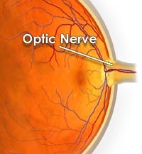
The Optic Nerve is located at the back of the eye. It transmits nerve imopulses from photoreceptors to the brain.
Blind Spot
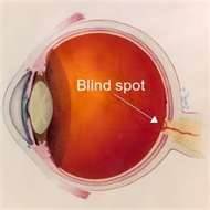
The point in the Retina where the optic nerve exits the eye. There are no photoreceptors which makes this Blind Spot insensitive to light.
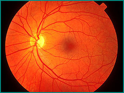
Fovea
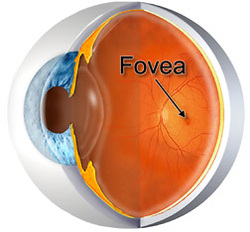
A pit in the Retina where images are focused. It has a high density of cones but it has no rods. The Fovea permits detailed vision. It is concentrated with photoreceptors.
